Tendon Diagram Labeled : 33 Muscular System Quiz Label Label Design Ideas 2020 : The patellar tendon holds the patella with other two bones, similarly iliotibial band helps in supporting tibia and fibula.
Tendon Diagram Labeled : 33 Muscular System Quiz Label Label Design Ideas 2020 : The patellar tendon holds the patella with other two bones, similarly iliotibial band helps in supporting tibia and fibula.. Rehabilitation of running biomechanics online course: The knee joint, you need a perfectly labeled diagram of the knee. Skin structure vector illustration diagram with skin layers and main elements. Can someone send me a link of the labellings of the hand. Reflex exam (deep tendon reflexes).
Rehabilitation of running biomechanics online course: Which of the labeled structures on the diagram holds muscles with similar functions together, allows free movement of muscles, carries nerves, blood vessels and lymphatic vessels, and. Have rich vascular supply and thus heal better. They have a central cell nucleus with a prominent nucleolus. Click here to download a free human skeleton diagram.
Human Muscle System Ankle Anatomy Muscle Anatomy Foot Anatomy from i.pinimg.com Understanding the structure of the foot is best done by looking at a foot diagram where the anatomy has been labeled. Tendons orient themselves along stress. Click here to learn the concepts of tendons from biology. Reflex exam (deep tendon reflexes). Rehabilitation of running biomechanics learn how to create a. Which of the labeled structures on the diagram holds muscles with similar functions together, allows free movement of muscles, carries nerves, blood vessels and lymphatic vessels, and. Now let's come to ligaments of the knee. Juan ramos on july 5, 2018 leave a comment!
Juan ramos on july 5, 2018 leave a comment!
Related online courses on physioplus. A tendon (or sinew) is a tough band of tissue that connects muscle to bone. If the tendon cannot be identified then a complete tear of the tendon should be sought. The reflex exam is fundamental to the neurological exam and important to locating upper versus lower motor neuron lesions. This tendon straightens the end joint of the thumb and also helps pull the thumb in towards the index finger. One is in color, and the other is in black and white. They have a central cell nucleus with a prominent nucleolus. Reflex exam (deep tendon reflexes). Medically reviewed by the healthline medical network — written by the healthline editorial team — updated on january 21, 2018. This entry was posted in anatomy by admin. I inhaled diatomaceous earth (labeled as silicon dioxide) to control insect issue at home, and have been coughing for two days with slight chest pain. answered by dr. Click here to download a free human skeleton diagram. Other tendon reflexes that could be examined are the triceps, supinator, ankle or achilles tendons and the babinski reflex.
Click here to learn the concepts of tendons from biology. The foot diagram has a complex structure made up of bones, ligaments, muscles, and tendons. The long extensor tendon to the thumb is called the extensor pollicis longus (epl). They have a central cell nucleus with a prominent nucleolus. The tendon runs around a bony prominence on the back of the wrist called lister's tubercle.
Ruptured Anterior Cruciate Ligament Racl Mar Vista Animal Medical Center from www.marvistavet.com A ruptured central slip tendon injury will cause the lateral bands to slide volarly and cause abnormal flexion of the pip joint and extension of the dip joint. The cytoplasm is stretched between the collagen fibres of the tendon. Includes labeled human skeleton chart. Rehabilitation of running biomechanics learn how to create a. The foot diagram has a complex structure made up of bones, ligaments, muscles, and tendons. One is in color, and the other is in black and white. Which of the labeled structures on the diagram holds muscles with similar functions together, allows free movement of muscles, carries nerves, blood vessels and lymphatic vessels, and. I inhaled diatomaceous earth (labeled as silicon dioxide) to control insect issue at home, and have been coughing for two days with slight chest pain. answered by dr.
Great for artists and students studying human anatomy.
The long head of biceps (lhb) tendon is usually located inferiorly in the bicipital groove held there by the biceps pulley (the stabilization role of th. The reflex exam is fundamental to the neurological exam and important to locating upper versus lower motor neuron lesions. Sgarlato (27) presented the flexor tendon transfer for. Click here to download a free human skeleton diagram. Now let's come to ligaments of the knee. Skin structure vector illustration diagram with skin layers and main elements. Understanding the structure of the foot is best done by looking at a foot diagram where the anatomy has been labeled. Labeled anatomy chart with two bones, articular cartilage, joint cavity, synovial fluid, muscle and tendon. A normal response involves an equal, appropriate response on both sides of the body. Which of the labeled structures on the diagram holds muscles with similar functions together, allows free movement of muscles, carries nerves, blood vessels and lymphatic vessels, and. This tendon straightens the end joint of the thumb and also helps pull the thumb in towards the index finger. One is in color, and the other is in black and white. Tendon cells, or tenocytes, are elongated fibroblast type cells.
Other tendon reflexes that could be examined are the triceps, supinator, ankle or achilles tendons and the babinski reflex. To understand one of the most complex joints of our body i.e. Muscle spindle and golgi tendon organs. They reported modifying the split principles of tendon surgery were then incorporated into the treatment of the equinus foot (25,26). Reflex exam (deep tendon reflexes).
ሠBack Muscle Diagrams Labeled Stock Vectors Royalty Free Trapezius Illustrations Download On Depositphotos from st2.depositphotos.com If the tendon cannot be identified then a complete tear of the tendon should be sought. Diatomite is an amorphous form of silica that is relative. Understanding the structure of the foot is best done by looking at a foot diagram where the anatomy has been labeled. A normal response involves an equal, appropriate response on both sides of the body. Medically reviewed by the healthline medical network — written by the healthline editorial team — updated on january 21, 2018. There are five deep tendon reflexes and a number of superficial and visceral reflexes covered here. The reflex exam is fundamental to the neurological exam and important to locating upper versus lower motor neuron lesions. This entry was posted in anatomy by admin.
Skin structure vector illustration diagram with skin layers and main elements.
Tendons, ligaments, bone, and cartilage are connective tissues in which the activities of various cellular populations are responsible for synthesis and maintenance of large amounts of extracellular matrix that should, theoretically, be dynamically optimized to respond to mechanical demands. One is in color, and the other is in black and white. The development of major tendon transfers in the podiatric community was first published by mcglamry (20) in 1973. Click here to download a free human skeleton diagram. There are 6 worksheets to choose from. The knee joint, you need a perfectly labeled diagram of the knee. The foot diagram has a complex structure made up of bones, ligaments, muscles, and tendons. Other tendon reflexes that could be examined are the triceps, supinator, ankle or achilles tendons and the babinski reflex. This entry was posted in anatomy by admin. Understanding the structure of the foot is best done by looking at a foot diagram where the anatomy has been labeled. A ruptured central slip tendon injury will cause the lateral bands to slide volarly and cause abnormal flexion of the pip joint and extension of the dip joint. The long head of biceps (lhb) tendon is usually located inferiorly in the bicipital groove held there by the biceps pulley (the stabilization role of th. If the tendon cannot be identified then a complete tear of the tendon should be sought.
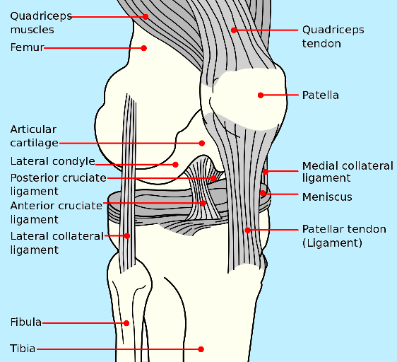
The knee joint, you need a perfectly labeled diagram of the knee tendon diagram. Tendons, ligaments, bone, and cartilage are connective tissues in which the activities of various cellular populations are responsible for synthesis and maintenance of large amounts of extracellular matrix that should, theoretically, be dynamically optimized to respond to mechanical demands.


0 Komentar